Fig. 2.
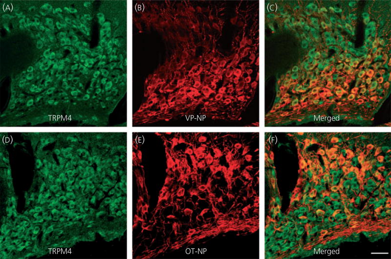
Confocal double immunofluorescence of TRPM4 with vasopressin (VP)-neurophysin (NP) or oxytocin (OT)-NP in the supraoptic nucleus (SON). (a, d) Confocal photomicrographs of TRPM4 immunoreactivity labelled with Alexa Fluor 568-conjugated secondary antibody in the SON. TRPM4 immunoreactivity was observed in most of magnocellular cell bodies in the SON. (b) VP-NP immunoreactivity labelled with Alexa Fluor 488-conjugated secondary antibody in the same optical section as in (a). (c) Merged image of (a) and (b). The TRPM4 immunoreactivity was co-localised with VP-NP immunoreactivity as indicated by yellow produced by an overlap of the Alexa Fluor 568 and 488 labelled elements (arrows). (e) Confocal photomicrographs of OT-NP immunoreactivity labelled with Alexa Fluor 488-conjugated secondary antibody in the same section and image plane as in (d). (f) Merged image of (d) and (e). The TRPM4 immunoreactivity was also co-localised with OT-NP within the cell bodies in the SON. Scale bar = 50 µm.

