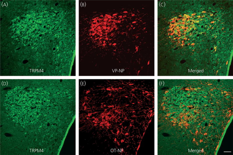
Fig. 3.
Confocal double immunofluorescence of TRPM4 with vasopressin (VP)-neurophysin (NP) or oxytocin (OT)-NP in the paraventricular nucleus (PVN). (a) Confocal photomicrographs of TRPM4 immunoreactivity labelled with Alexa Fluor 568-conjugated secondary antibody in the PVN. Prominent TRPM4 immunoreactivity is observed in somata of magnocellular cells (MNCs) within the PVN. Most of these TRPM4 immunoreactive MNCs are located in a cluster of cells in the lateral magnocellular region; however, TRPM4 immunoreactive magnocellular cells are scattered in other regions of the PVN (dorsal, medial, ventrolateral and posterior parvocellular regions). There are no prominent TRPM4 immunoreactivities among parvocellular cells in the PVN. (b) VP-NP immunoreactivity labelled with Alexa Fluor 488-conjugated secondary antibody in the same 2-µm optical section as in (a). (c) Merged image of (a) and (b). The TRPM4 immunoreactivity was co-localised with VP-NP immunoreactivity, as indicated by yellow colour produced by an overlap of the Alexa Fluor 568 and 488 labelled elements. (d) Confocal photomicrographs of TRPM4 immunoreactivity taken in the same optical section as in (e). (e) OT-NP immunoreactivity labelled with Alexa Fluor 488-conjugated secondary antibody. (f) Merged image of (d) and (e). As in the supraoptic nucleus, TRPM4 immunoreactivity was also co-localised with OT-NP. Scale bar = 50 µm.

