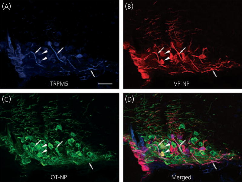
Fig. 5.
Confocal triple immunofluorescence of TRPM5, vasopressin (VP)-neurophysin (NP) and oxytocin (OT)-NP in the supraoptic nucleus (SON). (a) Confocal photomicrographs of TRPM5 immunoreactivity labelled with Alexa Fluor 647-conjugated secondary antibody in the SON. The most intense TRPM5 immunoreactivity was observed in the thick dendritic processes within the SON (arrows). Less intense but prominent TRPM5 immunoreactivity was also observed in the cell bodies. (b) VP-NP immunoreactivity labelled with Alexa Fluor 594-conjugated secondary antibody in the same optical section as in (a). (c) OT-NP immunoreactivity labelled with Alexa Fluor 488-conjugated secondary antibody in the same 2-µm optical section as in (a) and (b). (d) Merged image of (a), (b) and (c). TRPM5 immunoreactivities were co-localised well with those of VP-NP, and not OT-NP. These co-localisations are indicated by purple colour produced by overlap of the Alexa Fluor 647 and 594 labelled elements. Note there is no co-localisation of TRPM5 and OT-NP immunoreactivity either in the processes or cell bodies, except in rare cases where co-localisation of VP-NP and OT-NP immunoreactivites occured. These cases are indicated by the yellow colour and arrowheads. Scale bar = 50 µm.

