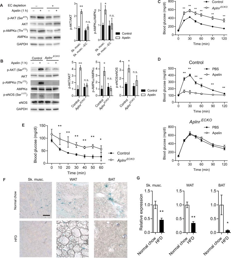
Fig. 2. Endothelial APLNR is critical for apelin signaling.
(A) Representative immunoblot of AKT and AMPKα phosphorylation in response to apelin stimulation in skeletal muscle with or without EC depletion (n = 3 replicates per group). GAPDH, glyceraldehyde-3-phosphate dehydrogenase; n.s., not significant. (B) Representative immunoblot of AKT, AMPKα, and eNOS phosphorylation in response to apelin stimulation in skeletal muscle of AplnrECKO mice and control littermates (n = 3 replicates per group). (C) IPGTT of AplnrECKO mice and control littermates [n = 10 (control) and 14 (AplnrECKO)]. (D) IPGTT in AplnrECKO and control mice 30 min after intravenous apelin injection [n = 5 (control) and 7 (AplnrECKO)]. (E) Intraperitoneal insulin tolerance testing of AplnrECKO mice and their control littermates under basal conditions (n = 6 per group). (F) Representative apelin expression in skeletal muscle, WAT, and BAT, as assessed by lacZ staining of Apln+/lacz reporter mice fed a normal chow or a high-fat diet (HFD). Scale bar, 100 µm. (G) Relative mRNA expression of apelin in various tissues of mice fed a normal chow or a high-fat diet (n = 5 per group). *P < 0.05 and **P < 0.01.

