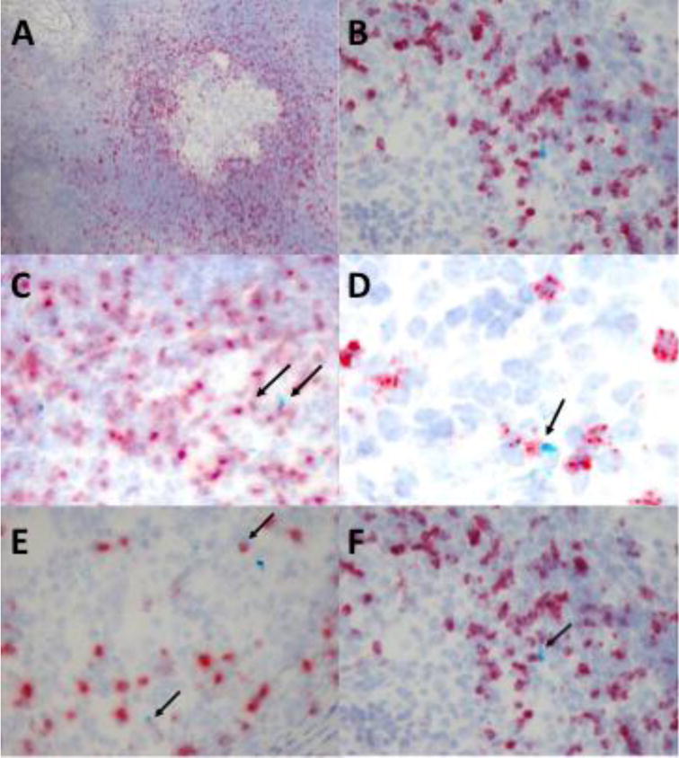
Figure 2. γδ T cells express various cytokines and chemokines within late-stage granulomas.

Tissue samples from granulomatous lesions in the lungs and mediastinal lymph nodes were harvested from calves (n=5) 3 months post-infection with virulent M. bovis. Tissue sections were preserved onto slides by formalin fixation and paraffin embedding. RNAScope was used for in situ analysis of mRNA transcripts of various cytokines/chemokines (turquoise) and the γδ TCR (red). Characteristic TB granuloma at 10X magnification (A) and at 40X magnification (B) within the lymph node of a chronically infected animal. Arrows indicate instances of γδ T cell and IFN-γ (C) IL-10 (D) CCL2 (E) IL-17 (F) co-expression.
