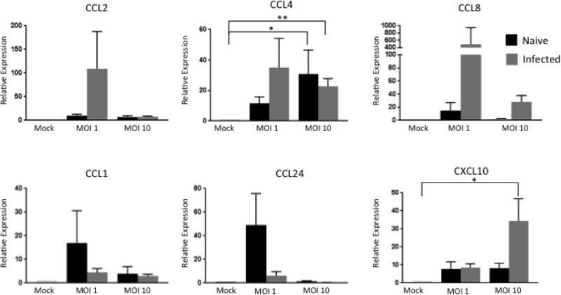
Figure 4. Chemokine expression in γδ T cell/MDM co-cultures from naïve and M. bovis-infected calves stimulated with BCG.

MDM and autologous γδ T cells isolated from M. bovis-infected or naïve animals were cultured together for 24 hours after a 4 hour infection with BCG at an MOI of 1 or 10. RNA was extracted and reverse transcribed into cDNA and qPCR was performed on various chemokines. Results were normalized to the housekeeping gene RPS-9, and expressed relative to uninfected γδ/MDM co-culture (mock) samples. Data represent means ± SEM (n=19 for naïve group and n=10 for infected group) (* P≤ 0.05; ** P≤0.01; ANOVA).
