
Figure 5. γδ T cells from M. bovis-infected calves are the main source of IL-17, IFN-γ and CXCL10 when in contact with BCG-infected MDM.
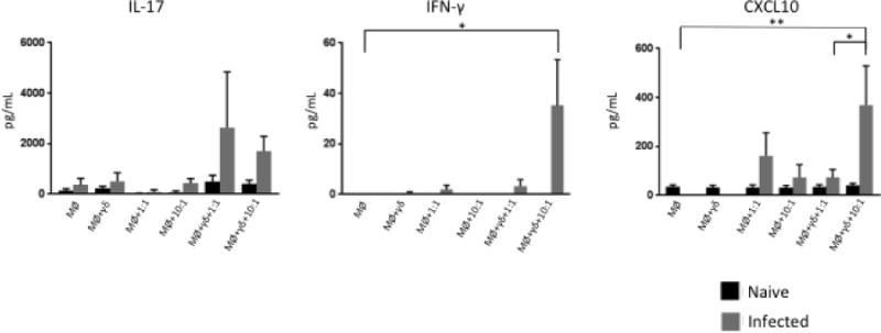
Commercial ELISA kits were used to measure IL-17A, IFN-γ, and CXCL10 from the supernatants of uninfected and BCG-infected co-cultures of MDM in direct contact with autologous γδ T cells. Data represent mean ± SEM (n=19 for naïve group and n=10 for infected group) (* P≤ 0.05; ** P≤0.01; ANOVA).