
Figure 1.
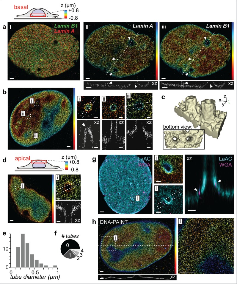
Investigating nanoscale nuclear envelope topography and invaginations by 3D SMLM. (a) A- and B-type lamins share the same topography. Lamin A and lamin B1 of fixed human foreskin fibroblasts were immunostained and imaged by dual-color 3D STORM at the basal side of the nucleus. Shown are (i) an overlay and (ii+iii) z-color coded images of individual lamins with line profiles underneath. Arrowheads indicate tube-like invaginations. Scale bars: 1 µm. (b) Tube-like invaginations at the basal side of a lamin A stained nucleus. Arrowhead: tip of an invagination. Scale bars: 1 µm (overview) or 0.2 µm (magnified insets i-iii). (c) 3D reconstruction of 2 nearby tubes. (d) Invagination at the apical side of a lamin A stained nucleus. Scale bars: 1 µm (overview) or 0.2 µm (inset i). (e) Pooled statistics of tube diameters (n = 57, from 30 cells). (f) Occurrence frequency of the number of tubes per imaged side of the nucleus (60 basal, 20 apical). As only one side could be imaged per nucleus, this represents a lower estimate. (g) Dual-color 3D STORM of A-type lamins (LaAc; cyan) and wheat germ agglutinin (WGA; magenta) as an NPC stain. Arrowheads: NPCs. Scale bars: 1 µm (overview) or 0.2 µm (insets).(h) 3D DNA-PAINT measurement of lamin B1. Scale bars: 1 µm.