
Figure 2.
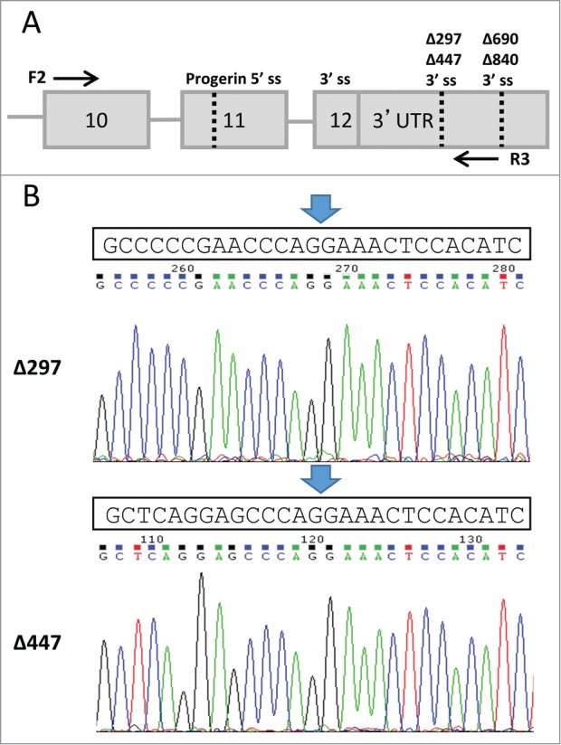
Confirmation of LMNAΔ297 and LMNAΔ447 isoform expression by RT-PCR. (A) Schematic representation of experimental setup for RT-PCR detection. Alternative 3′ splice acceptors and the progerin 5′ splice donor sites are indicated by dotted lines and labeled as splice site “ss." Flanking forward (F2) and reverse (R3) primers used for RT-PCR are shown as arrows. (B) Sequences of LMNAΔ297 and LMNAΔ447 were confirmed through gel extraction and direct sequencing of the PCR product. Predicted sequence is shown above the chromatograms for LMNAΔ297 and LMNΔ447, and the splice junction is indicated by arrow.