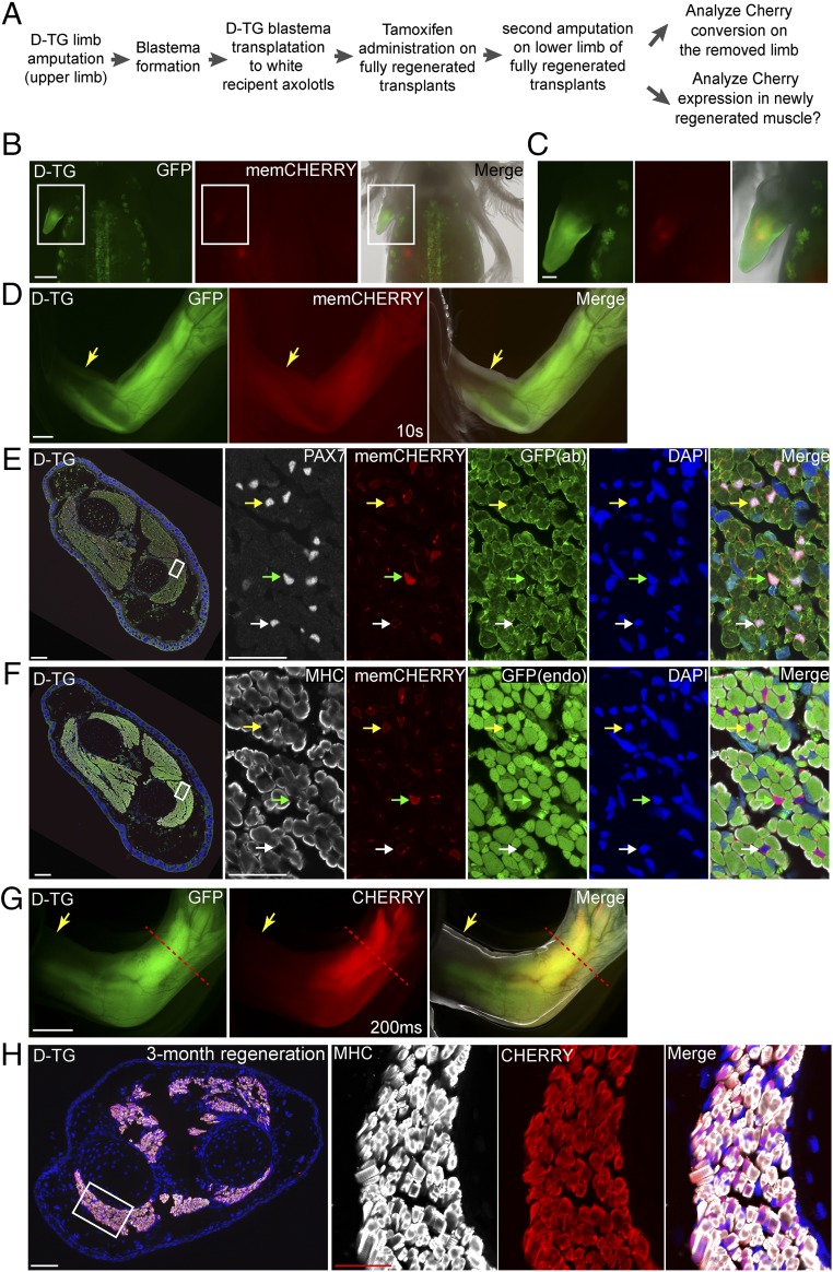
Fig. 3.
Lineage tracing of satellite cells during axolotl limb regeneration. (A) Scheme of the satellite cell lineage tracing experiment. (B–D) Live images of double-transgenic limbs after blastema transplantation and before tamoxifen treatment (n = 6). The GFP, CHERRY fluorescence, and merged (with bright-field) images of the transplanted limb blastema from the double-transgenic (D-TG) (SI Appendix, Fig. S7) donor to a white recipient at 2 wk (B and C) and 3 mo (D) post-transplantation. Note that the membrane-CHERRY signal is very dim in satellite cells in live imaging. Rectangular regions in B are shown at higher magnification in C. (Scale bars: 1 mm in B and D, 500 μm in C.) (E and F) Cross-section of limbs at 2 wk after tamoxifen treatment. Immunofluorescence for PAX7 (white, E) or MHC (white, F), CHERRY (red, memCHERRY), GFP [green, from antibody (ab) staining, in E] or endogenous (endo) GFP fluorescence (green, in F) combined with DAPI (blue) on adjacent 10-μm limb cross-cryosections (showing limb muscle) (n = 3). Green and yellow arrowheads indicate strong and dim cytoplasmic CHERRY-expressing (converted) cells, respectively. White arrowhead marks unconverted, memCHERRY-expressing cells. Muscle fibers (MHC-labeled) in the converted, mature limb remain CHERRY-negative. In E and F, the areas depicted by the rectangle in the leftmost panel are shown at higher magnification at the three right panels (Scale bars: 200 μm in left and 50 μm in right.) (G) Live images of tamoxifen-converted limbs after completion of regeneration: GFP, CHERRY fluorescence, and merged (with bright-field) images (n = 3). Arrows indicate the first amputation plane from the transplantation, and the red dashed line shows the plane of amputation after tamoxifen conversion. Exposure time for CHERRY, 200 ms. Note that Cherry driven by the CAGGS promoter is more strongly expressed in muscle tissues compared with satellite cells (10 s in Fig. 3D vs. 200 ms in Fig. 3G). Note that the GFP signal observed in the digit muscles represents GFP expression from incomplete conversion of the tandem array of LoxP reporter cassettes found in this reporter strain (SI Appendix, Fig. S10). (Scale bar: 1 mm.) (H) CHERRY-expressing satellite cells contribute to muscle regeneration. Cross-section of the limb that regenerated after induction of CHERRY expression from the LoxP reporter in PAX7-positive cells. Immunofluorescence for MHC (white) and CHERRY (red) combined with DAPI (blue) of a 3-mo regenerated limb showing robust expression of cytoplasmic CHERRY in muscle cells (n = 3). The area depicted by the rectangle in the leftmost panel is shown at higher magnification in the three right panels. (Scale bars: 200 μm in left and 50 μm in right.)

