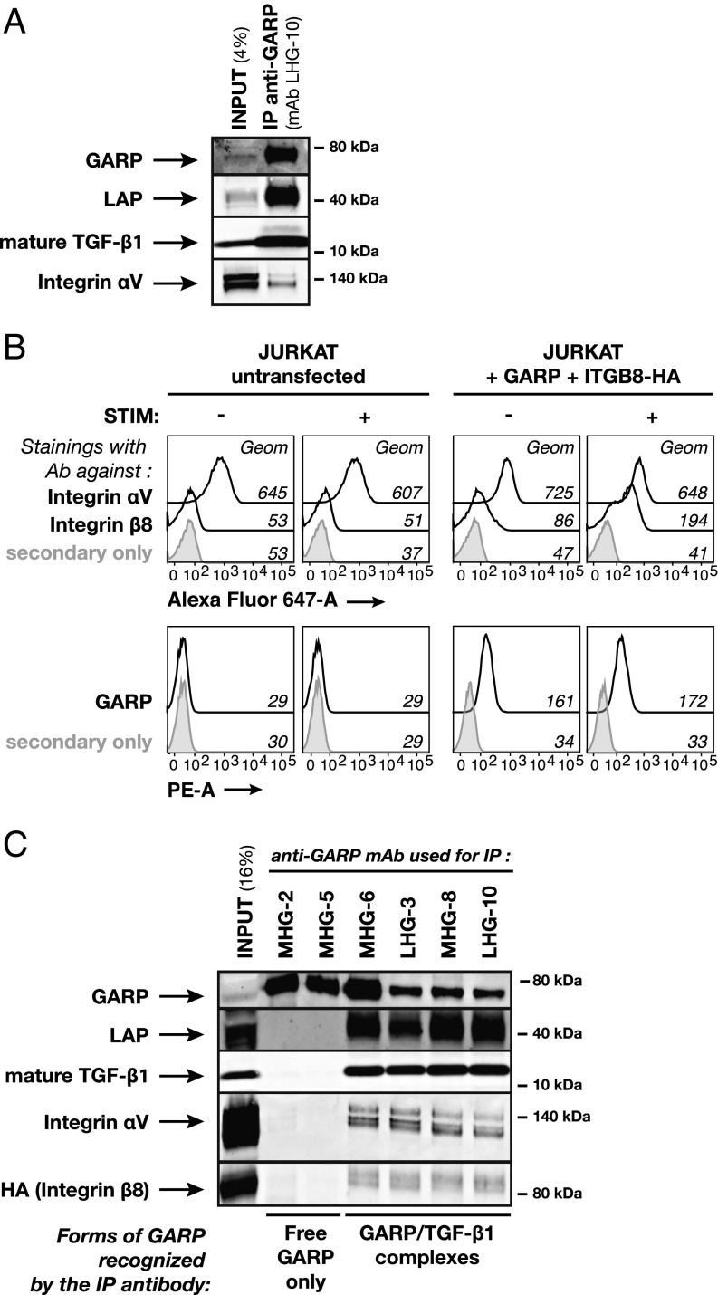
Fig. 4.
Integrin αVβ8 interacts with GARP/TGF-β1 complexes. (A) Polyclonal Tregs from donors 8 and 10 were stimulated with anti-CD3/CD28 antibodies for 24 h. Cell lysates were pooled and immunoprecipitated with anti-GARP mAb LHG-10. Total lysate (4% of input in IP) and IP product were analyzed by Western blot with antibodies against GARP, LAP, mature TGF-β1, or integrin αV subunit. Results are representative of three independent experiments. (B) Flow cytometry analyses of untransfected Jurkat cells (Left) or Jurkat cells transfected with GARP and HA-ITGB8 (Right), resting or 24 h after stimulation (STIM) with anti-CD3 antibody, and labeled with mAbs to integrin αV subunit or integrin β8 subunit followed by anti-mIgG1 antibodies coupled to Alexa Fluor 647 (Top) or with biotinylated anti-GARP mAb MHG-6 followed by streptavidin coupled to phycoerythrin (Bottom). (C) Jurkat cells transfected with GARP and HA-ITGB8 were stimulated with anti-CD3/CD28 antibodies for 24 h. Cell lysate was immunoprecipitated with the indicated anti-GARP mAbs. Total lysate (16% of input in IP) and IP products were analyzed by Western blot with antibodies against GARP, LAP, mature TGF-β1, integrin αV subunit, or HA (as a readout of integrin β8 subunit expression).

