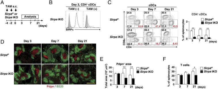
Fig. 3.
Importance of SIRPα on DCs for homeostasis of splenic FRCs in adulthood. (A) Sirpaf/f and Sirpa iKO mice were injected s.c. with TAM on days −4, −2, and 0. The spleen was isolated on days 3, 7, or 21 for immunohistofluorescence or flow cytometric analysis. (B) Staining for SIRPα (open traces) or with an isotype control antibody (filled traces) on CD4+ cDCs isolated from the spleens of Sirpa iKO mice 3 d after the last TAM [TAM(+)] or vehicle [TAM(−)] injection. Data are representative of three mice. (C) Representative flow cytometric profiles for CD8+, CD4+, and DN cDC subsets (Left) and the percentage of CD4+ cDCs (Right) in the spleens of Sirpaf/f or Sirpa iKO mice at 3, 7, and 21 d after the last TAM injection. (D) Frozen sections of spleens from Sirpaf/f and Sirpa iKO mice at 3, 7, and 21 d after the last TAM injection were stained with antibodies to Pdpn (red) and to B220 (green). (Scale bar, 500 μm.) (E) The Pdpn+ area in the spleens of Sirpaf/f and Sirpa iKO mice at 3, 7, and 21 d after the last TAM injection was measured in images similar to those in D with the use of ImageJ software. (F) The percentage of T cells among total splenocytes of Sirpaf/f and Sirpa iKO mice at 3, 7, and 21 d after the last TAM injection was determined by flow cytometry. All quantitative data (C, E, and F) are pooled from three independent experiments and represent the means ± SE for three mice per group. *P < 0.05, **P < 0.01 (Student’s t test).

