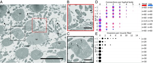
Fig. 3.
Fungal interactions observed in O. unilateralis s.l.-infected ant muscles. (A) Serial block-face SEM image showing fungal hyphal bodies (HB) and hyphae (arrowheads) occupying the spaces between ant mandible muscle fibers (M). Outlined boxes are shown larger in B and C. (Scale bar, 50 µm.) (B) Connections between hyphal bodies (arrows). (Scale bar, 10 µm.) (Inset) Close-up of connected hyphal bodies. (Scale bar, 1 µm.) (C) Muscle fiber invasion: hyphae have penetrated the membrane of this muscle fiber and are embedded within the muscle cell (arrows). (Scale bar, 10 µm.) (D) Connections per hyphal body across leg and head regions for eight ants. Point sizes are proportional to number of occurrences. “n” refers to number of hyphal bodies sampled in each ant. (E) Invasions per muscle fiber (head only) across eight ants (same ants as in D). “n” refers to the number of muscle fibers examined in each ant. Asterisks (*) denote ants that were dead at the time of collection. Images from A–C were all taken from Ant #1.

