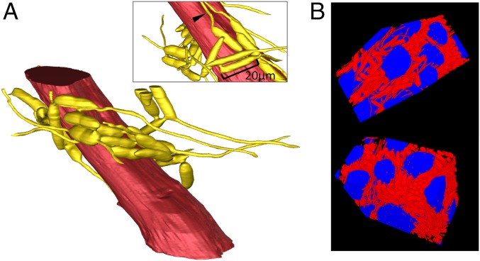
Fig. 4.
Three-dimensional reconstructions of fungal networks surrounding muscle fibers. (A) A single fiber of an ant mandible adductor muscle (red) surrounded by 25 connected hyphal bodies (yellow). Connections between cells are visible as short tubes, and many cells have hyphae growing from their ends. Some of these hyphae have grown along and parallel to the muscle fiber (arrowhead in Inset). This reconstruction was created using Avizo software. See also Movie S1 and interactive 3D pdf (Fig. S3). (B) Two different projections of a 3D reconstruction showing several muscle fibers (blue) and fungal hyphal bodies (red) from the same area as seen in A. This reconstruction was created using a method (developed here) that uses a U-Net deep-learning model.

