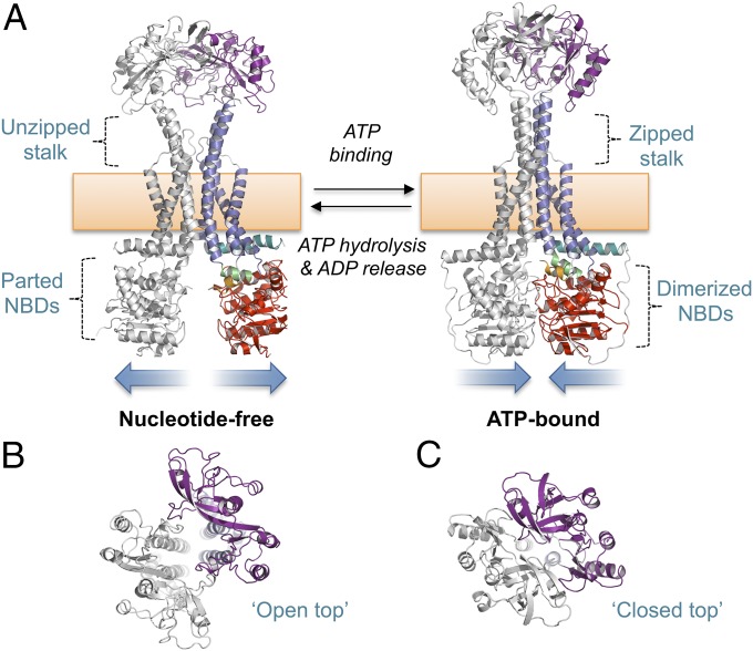
Fig. 4.
Mechanotransmission mechanism of MacB. (A) Nucleotide-free (Left) and ATP-bound MacB (Right). (B) Top-down view of the MacB dimer showing the open head of the nucleotide-free form. (C) Equivalent view of the ATP-bound form showing closure of the periplasmic dimer. Domains of MacB colored as in Fig. 1. Both models represent E. coli MacB; the nucleotide-free form is extracted from the cryoEM structure of the MacAB-TolC complex (5NIL), and the ATP-bound form is a homology model generated from our crystal structure of the nucleotide-bound AaMacB (5LIL). A full molecular morph between the two states is presented in Movie S3.

