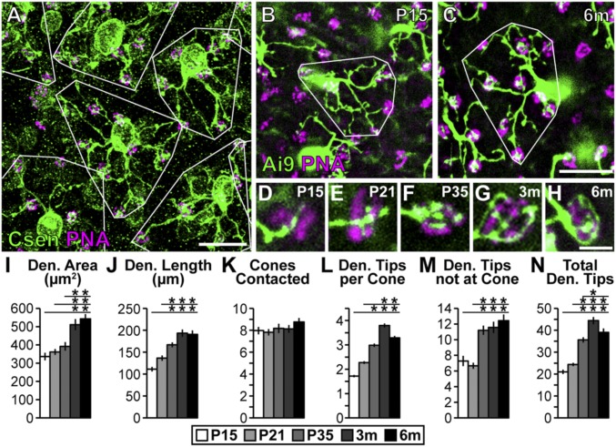
Fig. 1.
BC4s elaborate their dendritic arbors into adulthood. (A) Whole-mount retina (3 mo) stained with anticalsenilin antibody and PNA to visualize BC4s and cone photoreceptors, respectively. Dendrite areas are outlined in white to show tiling. (Scale bar, 25 μm.) (B and C) Genetically labeled BC4s (HTR:Cre × Ai9) at P15 (B) and 6 mo (C). Dendritic arbors occupy more area and elaborate their arbors throughout development and into adulthood. (Scale bar, 25 μm.) (D–H) High-magnification images of BC4 connections with cones from P15 to 6 mo. BC4s elaborate their innervation of cones over time until they become almost paddle shaped. (Scale bar, 5 μm.) (I–N) Quantification of BC4 characteristics at P15, P21, P35, 3 mo, and 6 mo. Significant increases in all measurements except number of cones contacted were detected (ANOVA P values in I, J, L–N: P < 0.0001; in K: P = 0.48). Horizontal lines above columns in I–N represent comparisons between the column under the left end of the line and all columns to the right. An asterisk above a point on the line represents a post hoc significance test value, indicating a significant difference between the left-most column under the line and the column under the asterisk. Traced cells: n = 36, 39, 40, 40, and 30 at P15, P21, P35, 3 mo, and 6 mo, respectively. Error bars = SEM. WT data included also in Figs. 2 and 3 and Figs. S4 and S5. Csen, calsenilin; Den., dendrite; m, month; P, postnatal day.

