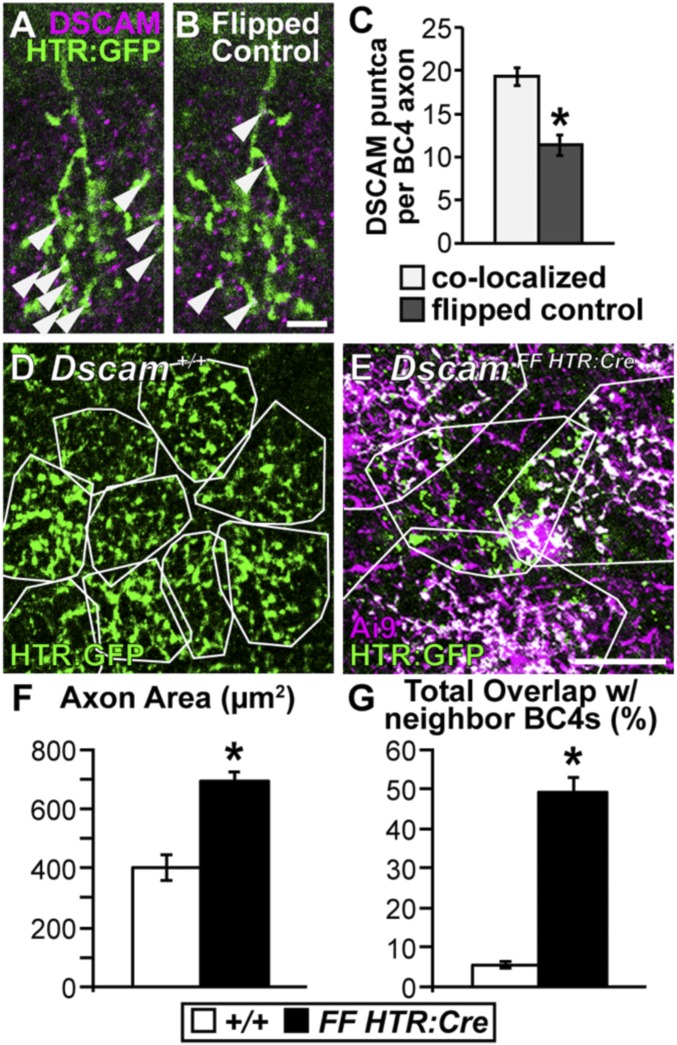
Fig. 7.
Dscam is necessary to establish BC4 axon tiling. (A–C) Colocalization of DSCAM protein with BC4 axons. All analyses were performed in serial z-stack images; shown here is a single confocal plane from an analyzed stack. (A and B) Cryo-sections of retina (3 mo) stained with anti-DSCAM and HTR:GFP to determine colocalization. (Scale bar, 5 μm.) (A) Image showing DSCAM/BC4 axon colocalization. (B) Axon channel flipped about the horizontal axis as a control for incidental colocalization. Arrowheads point to colocalized DSCAM puncta on BC4 axons. (C) Quantification of DSCAM puncta colocalization per BC4 axon. A significant increase in DSCAM puncta was detected when comparing the original images to the flipped controls (t test, P < 0.001). (D and E) Whole-mount retina (3 mo), BC4s are labeled genetically (HTR:Cre × Ai9 and HTR:GFP or HTR:GFP only). (Scale bar, 20 μm.) (D) Dscam+/+, BC4 axons outlined in white to visualize tiling. (E) DscamFF HTR:Cre, axons outlined in white. Only the green axon is Dscamflox. The others outlined are DscamΔflox. Axons invade each other’s territory when one or both lack DSCAM. (F and G) Quantifications of axon area (F) and total overlap with neighboring BC4 axons (G). Dscamflox BC4 axons become significantly larger (t test < 0.0001) and overlap significantly more with neighboring DscamΔflox BC4 axons (t test, P = <0.0001) compared with Dscam+/+ BC4 axons. *P < 0.05. Sample size WT, n = 30; Δflox, n = 30. Error bars = SEM.

