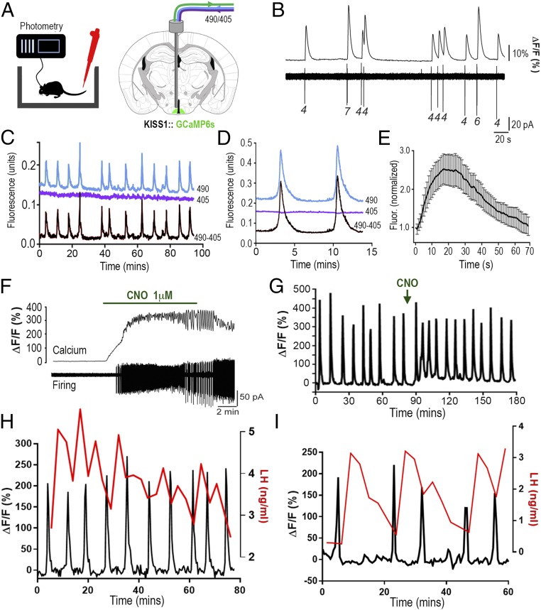
Fig. 1.
ARNKISS neurons exhibit periodic synchronizations in intracellular calcium concentrations tightly correlated with pulsatile LH secretion in gonadectomized male mice. (A) KISS1-Cre mice injected with Cre-dependent GCaMP6s AAV into the ARN (green) and connected to a fiber photometry system with regular blood sampling from the tail tip. Alternating 490-nm (Ca2+-dependent) and 405-nm (Ca2+-independent) wavelength light passes down the photometry fiber. (B) Simultaneous GCaMP6s calcium (Top) and cell-attached (Bottom) recordings from an ARNKISS neuron. Numbers indicate spikes per burst. (C) Fluorescence emission traces (490-nm calcium, blue; 405-nm background, purple) recorded from ARNKISS neurons of a gonadectomized male mouse with the subtracted signal (490 − 405) shown below. (D) Continuous recording of two calcium events from a gonadectomized mouse. (E) Average fluorescence waveform of calcium events (16 events, 7 mice) normalized to the beginning of each event. (F) Simultaneous brain slice recording of GCaMP6s fluorescence (Top) and action potential firing (Bottom) from a GCaMP6s/hM3Dq-expressing ARNKISS neuron showing the response to CNO applied in the bath. (G) Representative trace showing the effect of i.p. CNO (arrow) on GCaMP6 calcium signal from ARNKISS neurons in a gonadectomized mouse. (H and I) Representative dual calcium and plasma LH traces showing the near-perfect correlation of ARNKISS neuron GCaMP6 calcium events (black) with pulses of LH secretion (red) in two separate gonadectomized male mice. (I) The mouse with the slowest calcium events is shown.

