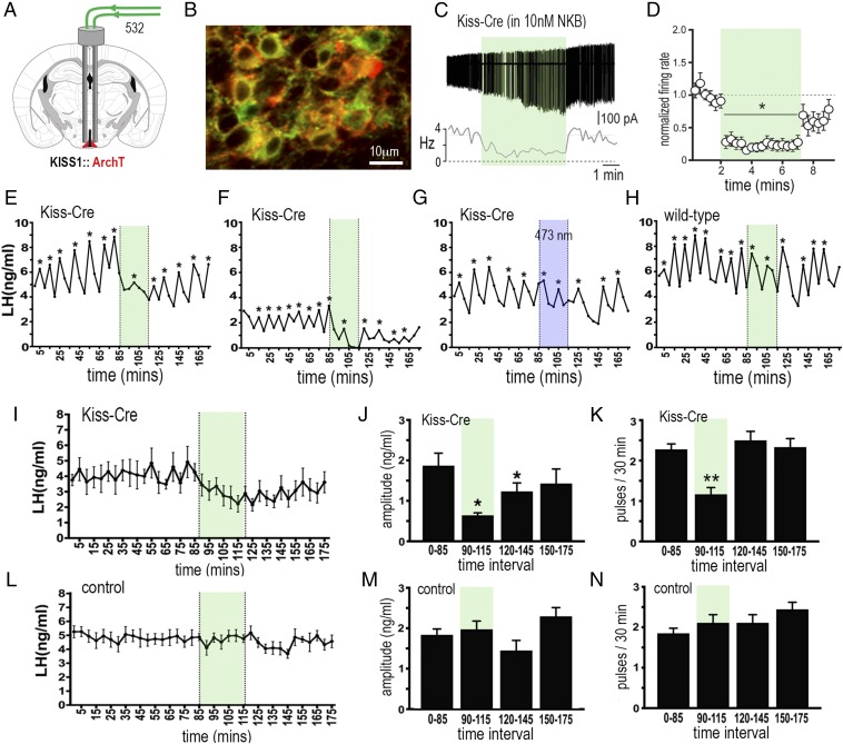
Fig. 3.
Archaerhodopsin inhibition of ARNKISS neurons suppresses pulsatile LH secretion in gonadectomized female mice. (A) KISS1-Cre mice injected with Cre-dependent ArchT-tdTomato AAV into the ARN (red) and implanted with bilateral optic fibers in the ARN. (B) Dual fluorescence image of ARNKISS neurons expressing ArchT (red) and kisspeptin (GFP reporter). (C) Action potential firing in an ArchT-expressing ARNKISS neuron in the presence of 10 nM NKB in a brain slice. The neuron responds to green light illumination (shading) with a decrease in firing followed by a return to control levels. (C, Top) Action potential firing. (C, Bottom) Rate meter for the same cell. (D) Mean (±SEM) normalized firing rate of ARNKISS neurons (n = 11) responding to green light. *P < 0.05 compared with baseline, Friedman test. (E–H) Pulsatile LH secretion in ArchT KISS1-Cre (E–G) and wild-type (H) gonadectomized female mice. Green light illumination (30 min) is indicated by green shading; LH pulses are indicated by asterisks. The trace in G shows the same mouse as in F but illuminated with blue light. (I) Mean (±SEM) LH levels in KISS1-Cre mice (n = 6) showing the suppression of LH secretion during green light illumination and subsequent slow recovery. (J and K) Mean (±SEM) LH pulse amplitude and pulse frequency before (0 to 85 min), during (90 to 115 min; green shading), and subsequent to (120 to 145 and 150 to 175 min) green laser illumination. *P < 0.05, **P < 0.01 versus 0 to 85 min, ANOVA with Dunnett’s post hoc tests; n = 6. (L–N) Basal (±SEM) LH levels and LH pulse amplitude and frequency in control mice including blue light-illuminated KISS1-Cre and wild-type AAV-injected mice (n = 8).

