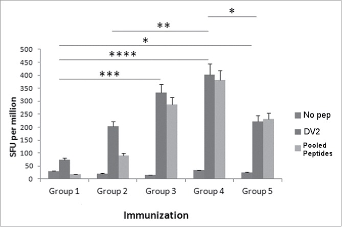
Figure 2.

Various concentrations of DV CaPNP/multipeptide formulations stimulate CD8+ T cell activation in vivo. HLA-A2+ transgenic mice were immunized as in Fig. 1A with the following groups: Group 1) unimmunized (PBS control); Group 2) 10µg peptide/150µl PBS emulsified in ISA 51 per mouse; Group 3) 50µg peptide/150µl PBS emulsified in ISA 51 per mouse; Group 4) 10µg peptide/150 µl CaPNP with 1XGlcNAc per mouse; Group 5) 50µg peptide/ 150 µl CaPNP with 1XGlcNAc per mouse. Splenocytes were harvested and co-cultured with HepG2 targets that were either pulsed with no peptides (negative control), pooled peptides (PP including NIQ, TIT, VTL, KLA, AML, LLC) or infected with DV2 for use in the ELISpot assay. Data represented as mean ± S.D (n = 3) of SFU per 1 million splenocytes. * Represents P values: * P<0.05, ** P<0.01, ***P<0.001, **** P<0.0001.
