Figure 4.

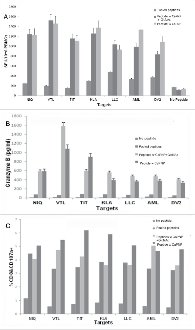
DV CaPNP/multipeptide formulations stimulate CD8+ T cell activation in vitro: (A) IFN-γ ELISpot assay: Using peripheral blood from healthy donors' PBMCs were stimulated in vitro with: 1) pooled peptides, or 2) CaPNP/multipeptide formulation with GlcNAc, or 3) CaPNP/multipeptide formulation without GlcNAc. The activated PBMCs containing the epitope specific CTLs were harvested, washed, and co-cultured overnight with HepG2 targets that were either pulsed with individual peptides (2µg each peptide/mL) or infected with DV2 in an IFN-γ ELISpot assay. Data represented as mean ± S.D (n = 3) of SFU per 1 million PBMCs. (B) Cytolytic response: Identical cultures were set up as in (A) in 96 well round bottom plates. The next day supernatant was harvested and used to detect cytolytic molecules (Granzyme B) using Luminex magnetic bead technology (MagPix). Data represented as mean ± S.D (n = 3) pg/mL. (C) Activation marker analysis: The cells were stained with CD8 and CD107a antibody at the end of the assay culture period to analyze the CTLs for the expression of degranulation marker, CD107a, by flow cytometry. Data represented as mean ± SD (n = 3) percent double positive (both CD8+ and CD107a+ staining) cells.
