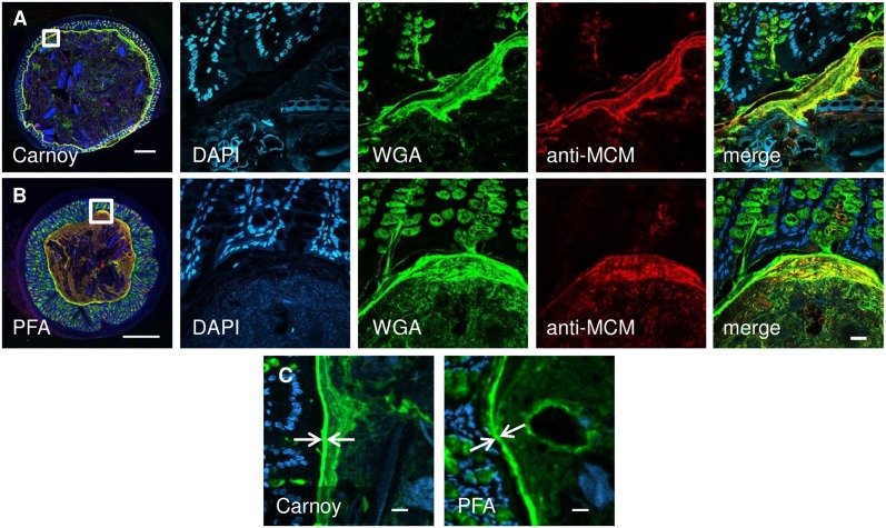
Fig 5. Carnoy and PFA fixation are both capable of preserving mucus, host tissue, and ingested food in intestinal segments that are embedded in methacrylate and sectioned.
Overview images (at left) show entire cross-sections of mouse proximal colon fixed with Carnoy (A) or paraformaldehyde (B). Middle panels show higher-magnification images stained with DAPI (blue), wheat germ agglutinin (green) and an antibody against mouse colonic mucin (anti-MCM, red) with a merged overlay image shown at right. Mucus, stained by both wheat germ agglutinin and anti-MCM, is preserved with both fixation methods. Detail images in (C) show the inner mucus layer (arrows) on which measurements were performed. Scale bar = 500 μm in overview images, 20 μm in other panels.

