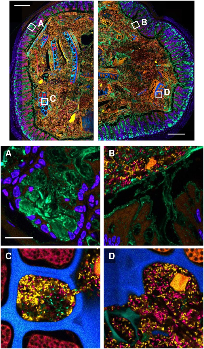
Fig 7. Visualization of the spatial organization of the gut environment using PFA fixation and methacrylate embedment.
A gnotobiotic mouse colon colonized with B. thetaiotaomicron and E. rectale was fixed with paraformaldehyde, embedded in methacrylate, sectioned, hybridized with oligonucleotide probes to differentiate the two microbial taxa, and stained with DAPI and with fluorophore-labeled wheat germ agglutinin. Fluorescence spectral images were coded to approximate true color. Top: tile-scanned overview images showing locations of detail panels (white boxes). Detail panels: (A): host cell nuclei (blue) and mucus in goblet cells (green). (B): the border between mucosa and lumen. Host cell nuclei are blue, mucus is green, B. theta is orange and E. rectale is red. Host tissue and a ten-micron-long food particle fluoresce orange. (C): a food particle with both colonized and un-colonized cavities. (D): a region of the lumen showing food particles with varying shapes and autofluorescence spectra in blue, green, and orange, signifying different types of food particles. Scale bars in overview images = 200 μm; scale bar in detail panel A = 20 μm and applies to all detail images.

