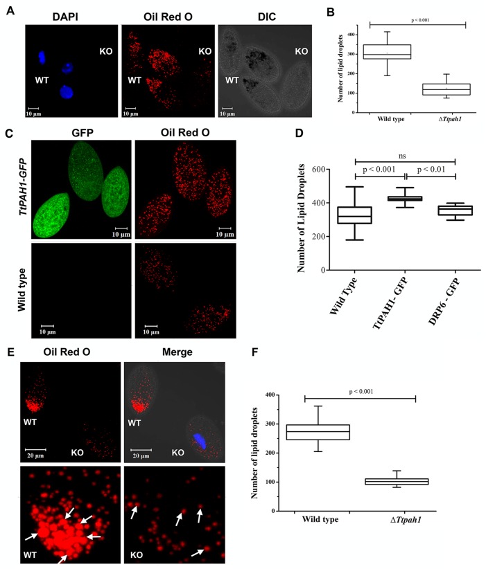
Fig. 4.
TtPAH1 maintains lipid droplet number in Tetrahymena. (A) Confocal images of Tetrahymena cells showing lipid droplets stained with Oil Red O dye. Wild-type cells and knockout cells were imaged together simultaneously. The wild-type cells were stained with DAPI to distinguish them from knockout cells. (B) Box plot showing the distribution of lipid droplet numbers in wild-type (n=35) versus ΔTtpah1 (n=38) cells. (C) Confocal images of wild-type and TtPAH1-GFP-expressing cells showing lipid droplets after staining with Oil Red O dye. (D) Box plot showing lipid droplet numbers in wild-type cells, cells overexpressing TtPAH1-GFP (n=20) and cells overexpressing GFP-DRP6 (n=20). An increase in lipid droplet number is observed in cells expressing TtPAH1-GFP. (E) Confocal images of Tetrahymena cells showing lipid droplets stained with Oil Red O dye. Wild-type (WT) and knockout cells after starvation were imaged together simultaneously. Knockout cells were stained with DAPI to distinguish them from wild-type cells. Both the size and number of lipid droplets are reduced in ΔTtpah1 cells (KO). Lipid droplet size in wild-type cells appears to be larger than in the knockout cells as indicated by arrows. (F) Box plot showing lipid droplet numbers in wild-type (n=22) and ΔTtpah1 (n=22) cells under starved condition.

