Figure 1.
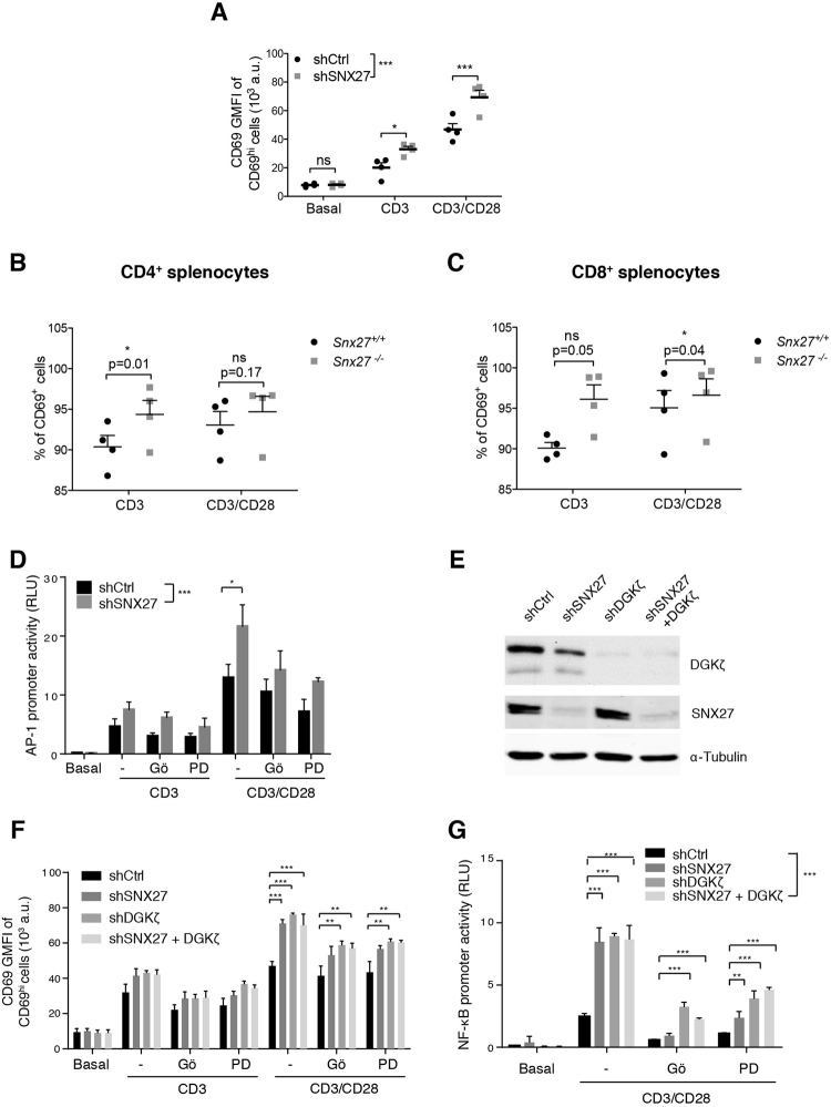
SNX27 limits activation of the Ras/ERK/AP-1 and NF-κB pathways. (A) shRNA-transfected Jurkat T cells were stimulated (6 h) with soluble anti-CD3 alone or with anti-CD28 for costimulation. Cells were stained for CD69 surface marker and, using flow cytometry, CD69hi cells were gated, and geometric mean fluorescence intensity (GMFI) of CD69hi cells was calculated. (B,C) Splenocytes from WT and Snx27 −/− littermate pairs were stimulated (48 h) with plate-bound anti-CD3 alone or with soluble anti-CD28 for costimulation and stained for CD69 surface marker. The percentage of CD69+ cells were gated in CD4+ (B) or CD8+ (C) cells. Data are shown as mean ± SEM (ns no significant, p > 0.05; *p < 0.05; paired t-test; n = 4). Luciferase assays were performed to calculate (D) AP-1 and (G) NF-κB promoter activity. RLU, relative luciferase units (see Methods). Where indicated, cells were pretreated with Gö6976 or PD98059 for classic PKC or MEK inhibition, respectively. (E) Western blot analysis of cell lysates from shControl and shSNX27/DGKζ Jurkat T cells (F) shControl and shSNX27/DGKζ Jurkat T cells were treated as in A with or without inhibitors where indicated. Two-way ANOVA with the Bonferroni post-hoc test was used for multiple comparisons, both with GraphPad Prism 5 software. (ns) when p > 0.05, (*) when p < 0.05, (**) when p < 0.01, (***) when p < 0.001.

