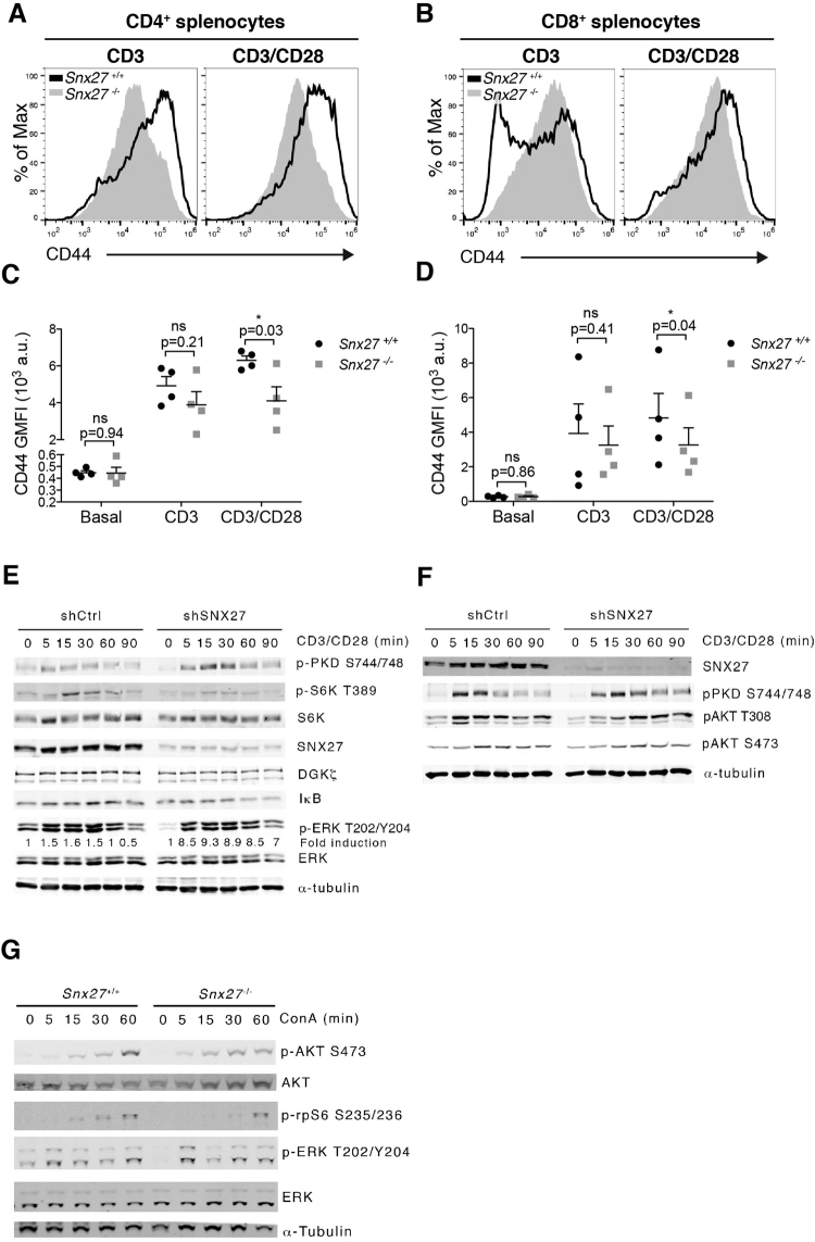
Figure 5.
Effect of SNX27 silencing on TCR/CD28 triggered signals. (A,B) Splenocytes from WT and Snx27 −/− littermate pairs were stimulated (48 h) with plate-bound anti-CD3 alone or with soluble anti-CD28 for costimulation and stained for CD44 surface marker. Representative flow cytometry plots for CD44 induction are shown. (C,D) CD44 GMFI were calculated. Data are shown as mean ± SEM (ns no significant, p > 0.05; *p < 0.05; paired t-test; n = 4) a.u., arbitrary units. (E,F) Control and SNX27-silenced cells were stimulated with anti-CD3/anti-CD28 antibodies for the indicated times. Western blot analysis of cell lysates showed phospho- and total protein abundance of the indicated proteins. Phospho-ERK signals were normalized to total ERK abundance, and results for each time point were also normalized to unstimulated cells to calculate fold induction. Blots are representative experiments from three independent experiments that provided similar results. (G) Splenocytes from WT and Snx27 −/− littermate pair were stimulated at the indicated times with concanavalin A (ConA). Phosphorylation of the indicated proteins was evaluated by western blot in total cell lysates. Tubulin was used as a loading control. A representative experiment from three independent ones is shown.

