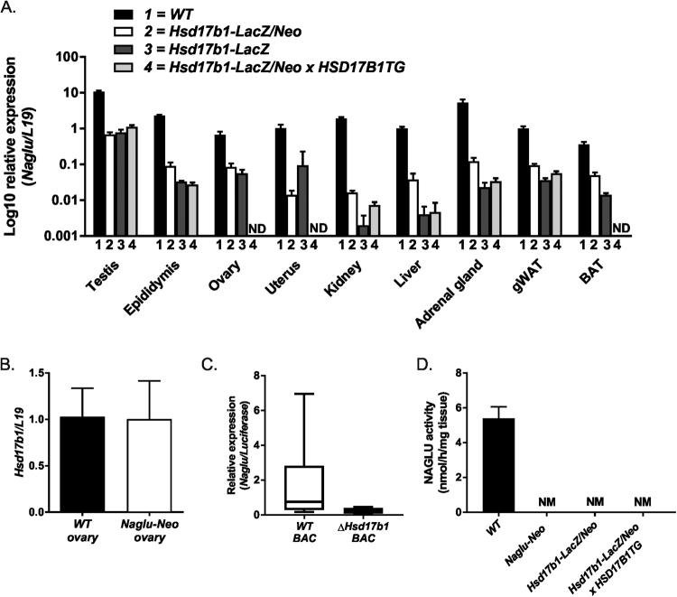
Figure 2.
Expression analysis of Naglu and Hsd17b1. Quantitative RT-PCR (A) analysis of the testis, epididymis, ovary, uterus, kidney, liver, adrenal gland, gonadal white adipose tissue (gWAT) and brown adipose tissue (BAT) showed the downregulation of Naglu expression in the Hsd17b1-LacZ/Neo mice (white bars), Hsd17b1-LacZ mice (dark grey bars) and Hsd17b1-LacZ/Neo X HSD17B1TG mice (light grey bars) compared with WT mice (black bars) at 3 months of age. (B) The Hsd17b1 mRNA expression was not altered in Naglu-Neo mice ovaries. Bars demonstrate the mean of the relative expression normalised to the expression of L19 (the error bars show SD, n = 6). (C) The expression of Naglu was downregulated in the BAC vector with the entire Hsd17b1 gene deleted (expression normalised to luciferase gene expression and error bars show SEM, n = 6). (D) Naglu activity was proven to be disrupted in the livers of Naglu-Neo, Hsd17b1-LacZ/Neo and Hsd17b1-LacZ/Neo X HSD17B1TG mice. Bars demonstrate the mean activity (Error bars SEM, n = 3–5). ND = not determined, NM = not measurable.

