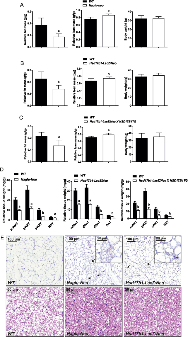
Figure 5.
Measurement of the fat and lean mass in the Naglu-Neo, Hsd17b1-LacZ/Neo and Hsd17b1-LacZ/Neo X HSD17B1TG male mice. (A) The fat mass was markedly reduced, and the lean mass was significantly increased in the Naglu-Neo mice (n = 9–10), while the body weight was not changed. Similar alterations were also observed in the Hsd17b1-LacZ/Neo (n = 10–11) (B) and Hsd17b1-LacZ/Neo X HSD17B1TG mice (n = 5–6) (C). (D) The weight of all the white adipose depots (scWAT = subcutaneous white adipose tissue, gWAT = gonadal white adipose tissue and pWAT = perirenal white adipose tissue), along with the brown adipose tissue (BAT), were decreased significantly. Statistical significance is marked with letters indicating P values: a, p < 0.001 compared to WT; b, p < 0.01 compared to WT; c, p < 0.05 compared to WT. Bars show mean value and error bars show SD. (E) Histological analysis showed smaller adipocytes in the WAT and the lipid droplets in BAT were smaller in size. Furthermore, the subcutaneous WAT of the Naglu-Neo and Hsd17b1-LacZ/Neo mice showed signs of browning (arrows), which is shown more detailed in insertions.

