Figure 2.
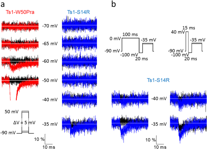
Voltage dependence of the ionic currents near threshold in wild-type Nav1.4 and in presence of Ts1-W50Pra or Ts1-S14R. (a) Ionic currents before applying toxin (black traces) and in the presence of toxin (Ts1-W50Pra in red and Ts1-S14R in blue). The traces in each column were obtained from the same cell before and after applying 1 µM of toxin and were superimposed after normalization by peak current. The inset in the lower-left part shows the protocol used. Scale bars are amplitudes (10% of peak current) and time (10 ms). (b) Currents before toxin (black traces) and in presence of Ts1-S14R when depolarized conditioning prepulse protocols were applied. These data was obtained from the same cell shown on the right column in (a). The protocols used are shown at the top. Current traces were obtained from the same oocyte. Scale bars, as in part a.

