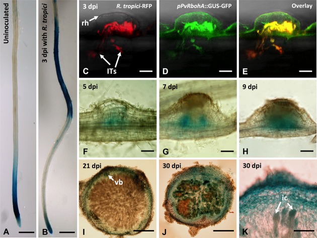
FIGURE 3.
Promoter analysis of PvRbohA in transgenic P. vulgaris roots and nodules. Spatiotemporal pattern of PvRbohA expression revealed by a promoter::GUS-GFP construct in 10-day-old transgenic hairy roots incubated with GUS substrate. (A) Uninoculated root. (B) R. tropici-inoculated root at 3 dpi. Confocal microscopy imaging of PvRbohA promoter activity in Rhizobium-infected root hair cell and in growing ITs at 3 dpi. (C) Red fluorescence emitted by R. tropici CIAT899 Ds-Red. (D) PvRbohA expression revealed by promoter::GUS-GFP and (E) overlay. PvRbohA promoter activity in the developing nodules at (F) 5 dpi, (G) 7 dpi, and (H) 9 dpi. (I) Mature 21-dpi nodule section showing PvRbohA promoter activity restricted to the nodule cortex and vascular bundles (vb). (J) With the onset of nodule senescence (30 dpi), the promoter was active in the central tissue containing infected cells. (K) Higher magnification of a senescing nodule section showing promoter::GUS-GFP activity in infected cells. rh, root hair; ITs, infection threads; ic, infected cell; hpi, hours post-inoculation; dpi, days post-inoculation. Bars: (A,B) 1 mm; (C–E) 10 μm; (F,G) 100 μm; (H,I) 200 μm; (J) 150 μm; (K) 50 μm.

