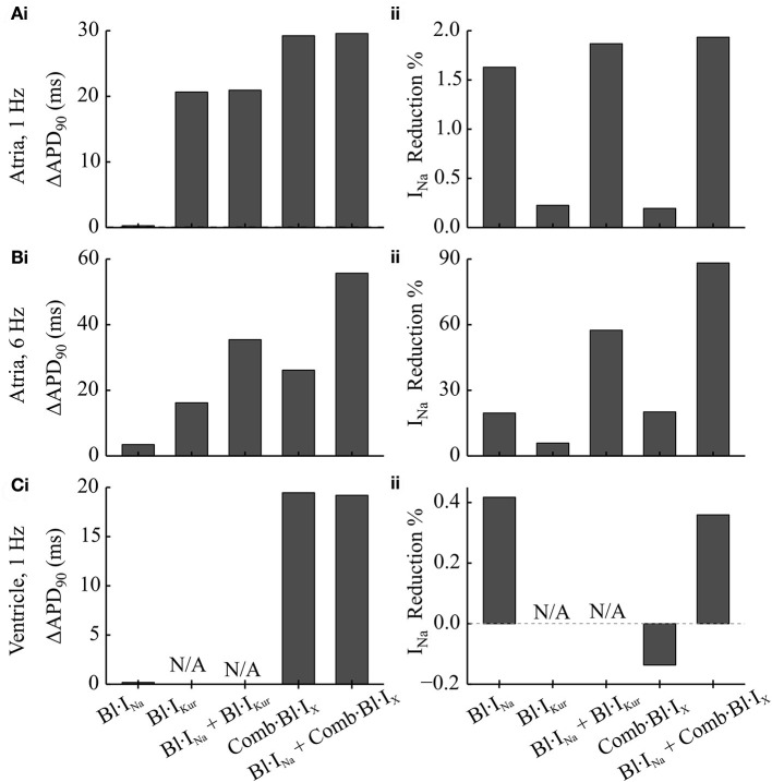
Figure 4.
Simulated changes in the APD and peak INa following the applications of Na+- and K+- block in comparison to the drug-free condition in cAF-remodeled atrial myocytes or ventricular cells. (A,B) Changes in (i) APD and (ii) peak INa measured from a cAF-remodeled atrial myocyte paced at (A) 1 Hz and (B) 6 Hz. In the presence of alternans, the changes in APD were quantified by comparing the corresponding longer APs. The fractional reductions in peak INa were calculated from the INa of the corresponding shorter APs. (C) Changes in (i) APD and (ii) peak INa measured from an in silico ventricular myocyte paced at 1 Hz.

