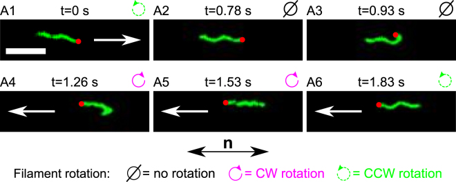
Figure 3.
Sequence of images taken from movie S2A (see also movies S2 B and S2C) showing an example of the locked hook during a reversal of the filament (reorientation of 180°). Above each image, we show the step label corresponding to the schematic drawing in Fig. 5, the time stamp of each movie frame, and the direction of rotation of the filament. The white arrows indicate the direction of the displacement of the bacterium (the mean speed of single flagellated bacteria was 1.7 ± 0.9 μm/s). There is no arrow in A2 and A3 because the cell body is at rest. The red dot identifies the base of the filament. The white scale bar at the bottom left of the first image measures 5 μm. The bottom arrow labeled n indicates the orientation of the anisotropy axis of the LC (identical in all frames).

