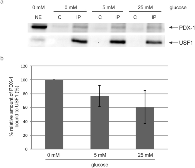
Figure 2.

Co-immunoprecipitation of PDX-1 and USF1. (a) INS-1 cells were seeded on a 14.5 cm culture plate and starved overnight. The next day, cells were treated with 0 mM, 5 mM or 25 mM glucose and after a period of 4 h cytoplasmic and nuclear proteins were extracted as described in material and methods. Two mg nuclear extract were incubated with the USF1 specific antibody sc-8983 for 2 h, immunocomplexes were loaded on a 10% SDS polyacrylamide gel and transferred onto a PVDF membrane. PDX-1 was identified with the polyclonal rabbit antiserum against recombinant full-length mouse PDX-1, detection of USF1 with the USF1 specific antibody sc-8983 served as a control for immunoprecipitation. NE: 50 μg nuclear extract; C: control precipitate; IP: immunoprecipitate. (b) Relative protein amounts of bound PDX-1 to USF1 were shown in the bar graphs. The diagram shows the mean ± SD of three independent experiments. The relative intensities of bound PDX-1 protein to USF1 were normalized to the protein content of precipitated USF1. Full-length blots are presented in Supplementary Figure S2.
