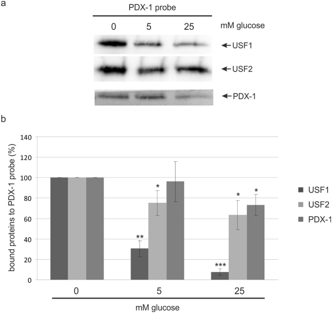
Figure 4.

Pull-down assay with the PDX-1 promoter in INS-1 cells treated with glucose. One mg of nuclear extract from INS-1 cells, who had been treated with 0 mM, 5 mM or 25 mM glucose for 4 h, was incubated with 1 μg of a biotinylated PDX-1 DNA probe. The DNA-protein complex was passed through a μMacs™-column and loaded on a 10% SDS polyacrylamide gel for Western blot analysis. (a) Identification of USF-binding at the PDX-1 promoter was performed with the USF1 specific antibody sc-8983 and with the USF2 specific antibody sc-862. PDX-1 was visualized with the polyclonal rabbit antiserum against recombinant full-length mouse PDX-1. Full-length blots are presented in Supplementary Figure S2. (b) Relative protein amounts of USF1, USF2 and PDX-1 bound to the PDX-1 promoter DNA. The diagram shows the mean ± SD of three independent experiments. Statistical analysis was performed by using Students t-test. *Significant difference p < 0.05, **significant difference p < 0.01, ***significant difference p < 0.001.
