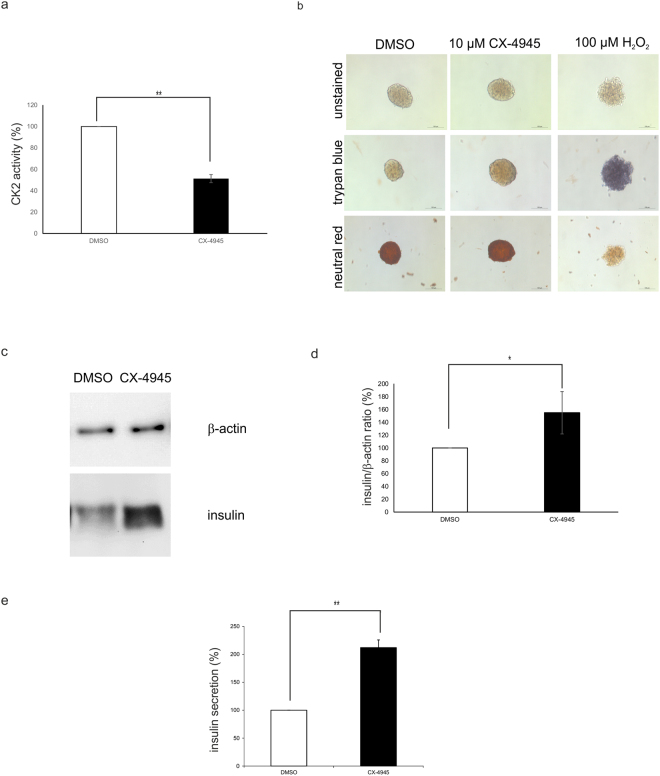
Figure 8.
Effect of CK2 inhibition on insulin expression and secretion of isolated pancreatic islets. (a) Isolated islets were treated with CX-4945 (10 µM) or DMSO as a solvent control for 24 h. Subsequently, protein extracts were generated and CK2 kinase activity was determined. DMSO treated islets were set 100%. Statistical analysis was performed by using Students t-test, **p < 0.01. (b) Light microscopic images of isolated islets which were treated with CX-4945 (10 µM) or DMSO as a solvent control for 24 h. Treatment with 100 µM H2O2 served as positive control for a treatment with an impact on cell viability. After treatment, islets viability was analysed after trypan blue and neutral red staining with a LEICA DMIL microscope and a LEICA DFC450 C camera. Scale bars: 100 μm. (c) Isolated islets were treated with CX-4945 (10 µM) or DMSO as a solvent control for 24 h and the expression of insulin and β-actin was analysed by Western blot. Full-length blots are presented in Supplementary Figure S2. (d) Quantitative analysis of (c). DMSO-treated islets were used as control and set 100%. Statistical analysis was performed by using Students t-test, *p < 0.05. (e) Pancreatic islets were treated with DMSO or 10 µM CX-4945 for 18 hours. After a glucose stimulus secreted insulin was determined in the cell culture supernatant with the rat/mouse insulin ELISA kit. DMSO treated islets were set 100%. Statistical analysis was performed by using Students t-test, **p < 0.01.

