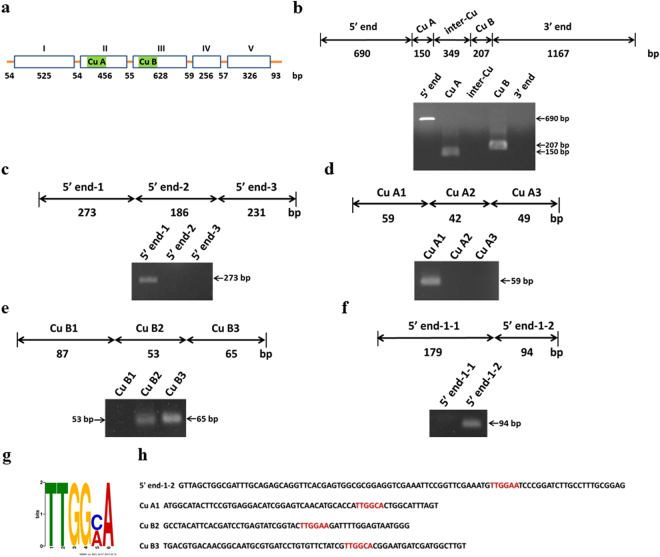
Figure 7.
Aacec B peptide binds to the putative binding motif TTGG(A/C)A present in four DNA fragments of AaPPO 3. (a) A schematic representation of the AaPPO 3 gene. Open boxes with roman numerals indicate exons and the nucleotide length of introns and exons are indicated by numbers. The green boxes indicate the copper binding site Cu A and Cu B. (b–f) DNA pull-down assay showing Aacec B peptide binding to AaPPO 3 DNA. The upper figures show schematic representations of the AaPPO 3 DNA fragments. The nucleotide lengths are indicated by numbers. The lower figures are the PCR analyses of the AaPPO 3 DNA fragments that are able to bind to Aacec B-conjugated beads. The arrows point to the PCR amplifications of the AaPPO 3 DNA fragments binding to the Aacec B-conjugated beads. (g) DNA sequence logo representing the TTGG(A or C)A binding motif that was identified from the four AaPPO 3 DNA fragments (5′ end-1-2, Cu A1, Cu B2 and Cu B3) using the MEME suite. The height of each letter represents the relative frequency of occurrence of the nucleotide at each position. (h) The nucleotide sequences of the four AaPPO 3 DNA fragments (5′ end-1-2, Cu A1, Cu B2 and Cu B3) containing the TTGGAA and TTGGCA putative motifs (indicated in red). Uncropped images are shown in Supplementary Figure S5.

