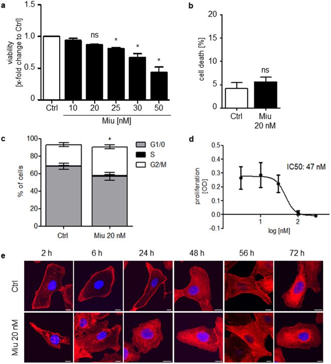
Figure 1.
Low dose treatment of SKOV3 with miuraenamide A (Miu) showed no effects on cell viability, proliferation and actin cytoskeleton morphology. (a) Cell viability after treatment for 72 h of SKOV3. (b) PI exclusion assay after treatment for 72 h with 20 nM Miu. (c) Cell cycle analysis. (d) Proliferation after treatment with increasing concentrations of Miu. (e) Actin staining of SKOV3 cells treated with 20 nM Miu for the indicated time points (rhodamine-phalloidin, red, and nucleus, Hoechst 33342, blue). White bars: 10 µm. ns, not significant. One-Way ANOVA/Bonferroni’s Multiple Comparison Test, *p < 0.05. n = 3.

