
Figure 1.
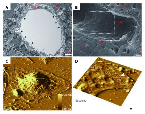
Morphological changes of liver sinusoidal endothelial cells. A: TEM image of rat liver sinusoid after transversal cut; B: SEM image of rat liver sinusoid; C: AFM image of LSECs with a 10 nm gold layer; D: A high scan range of AFM image of LSECs. A-B: ref 21 Copyright @1985 by the American Association for the Study of Liver Diseases; C-D: ref 22 Copyright @ 2012 Elsevier Ltd. AFM: Atomic force microscopy; LSECs: Liver sinusoidal endothelial cells; SEM: Scanning electron microscopy; TEM: Transmission electron microscopy.