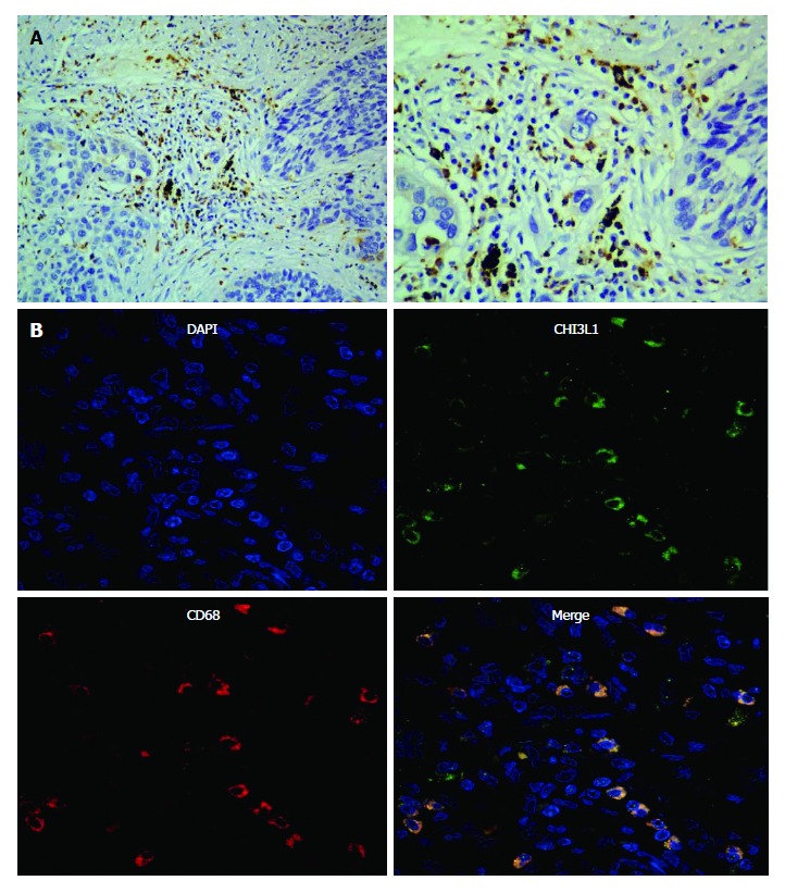
Figure 2.

Representative micrographs of esophageal squamous cell carcinoma tissues after immunohistochemistry and immunofluorescence assay. A: Sections stained with a CHI3L1 specific antibody (left- × 200; right- × 40); B: Sections immunofluorescence stained with CHI3L1 (upper left), an antibody against the macrophage specific marker CD68 (upper right), a nuclear counterstain DAPI (lower left) and their merge (lower right). CHI3L1: Chitinase 3-like 1.
