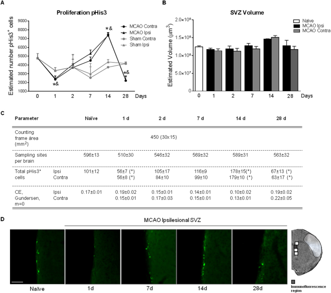
Figure 2.
Effect of stroke on SVZ proliferation analyzed by pHis3+ immunofluorescence in (A) MCAO (n = 6–8), sham (n = 4) and naïve (n = 8) animals. (B) Estimated SVZ volume calculated by Cavalieri estimator. (C) Parameters used for the stereological quantification of pHis3+ cells. (D) Representative images of pHis3 immunohistochemistry of naïve and ischemic groups at different time points after experimental ischemia (scale bar: 50 µm). Naïve group was represented in figure as t = 0. No differences were observed when naïve and sham animals were compared. Data are expressed as mean +/− SEM and compared by non-parametric 2-way ANOVA followed by Bonferroni post-hoc testing (F(1,38) = 12.26, *p < 0.0012, Naïve vs. MCAO 1d; F(1,20) = 7.642, &p = 0.0120, Sham vs. MCAO 1d; F(1,32) = 17.32; *p = 0.0002, Naïve vs. MCAO 14d; F(1,10) = 170.1, &p < 0.0001, Sham vs. MCAO 14d; F(1,38) = 12.21; *p = 0.0012, Naïve vs. MCAO 28d; F(1,16) = 12.01, &p = 0.0032, Sham vs. MCAO 28d).

