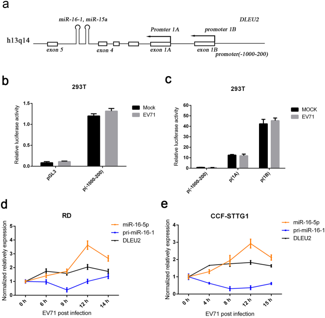
Figure 2.
EV71 promotes pri-miR-16-1 processing. (a) Schematic of the miR-16-1 gene and its promoter regions. Promoter 1 A and promoter 1B are two previously reported alternative promoters. The promoter (−1000–200) was from 1000 bp upstream to 200 bp downstream of oriented transcriptional start sites. (b,c) The EV71infection does not influence the transcription of pri-miR-16-1. Promoters 1A and 1B and −1000–100 regions were cloned to pGL3 vectors. Transcription experiments were performed with indicated reports or empty vectors together with an internal control, pRL-TK. Transfected 293 T cells were subjected to EV71 or mock infection for another 12 h. Then, reporter activity was determined by dual luciferase reporter assays. (d,e) Expression analysis of pri-miR-16-1 and DLEU2 in EV71-infected RD (d) and CCF-STTG1 cells (e). After EV71 virus infection for the indicated time, the expression levels of pri-miR-16-1 and DLEU2 were quantified by using qRT-PCR and the expression level of GAPDH as internal control. The relative expression levels of the target genes were calculated according to the 2−∆∆Ct method. Data are representative of a minimum of three independent experiments, with each determination performed in triplicate.

