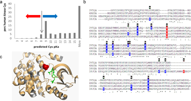
Figure 3.
Cysteine modification in human kinases. (a) Predicted cysteine pKas for a non-redundant set of human protein kinases, with red and blue arrows marking pKa decreased or increased, respectively, from the model compound pKa of cysteine (8.3). (b) Kinase domains of 4 kinases (3v5l A, 4rx5 A, 3pozA, 3pjcA) are aligned, with colour coding of cysteines according to a predicted lowering (red, more reactive) or raising (blue) of the sidechain pKa. Accessibility of each cysteine is marked as ‘a’ (accessible) or ‘b’ (buried). (c) EGFR kinase domain is shown for 3poz chain A, with inhibitor (green), and reactive cysteine site (red) at an α-helix amino-terminus.

