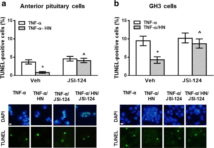
Fig. 2.
HN protected anterior pituitary cells and GH3 cells from TNF-α-induced apoptosis through STAT3 activation. (a) Anterior pituitary cells from OVX rats cultured with 17β-estradiol (E2, 10−9 M) or (b) GH3 cells were incubated with STAT3 inhibitor (JSI-124, 1 μM) for 30 min before adding HN (0.5 μM) for 2 h and TNF-α (50 ng/ml) for a further 24 h. Apoptosis was assessed by TUNEL. Each column represents the percentage ± CL of TUNEL-positive cells (n ≥ 1000 cell/group). * p < 0.05 vs respective control without HN, ^ p < 0.05 vs respective control without STAT3 inhibitor. χ2 test. The lower panels show representative images of TNF-α-induced apoptosis in anterior pituitary cells or GH3 cells incubated with HN in presence of STAT3 inhibitor. Scale bars: 10 μm

