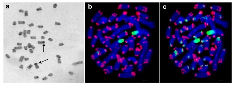
Figure 1.
Metaphase plates of female embryo of Medauroidea extradentata. (a) C-banding: large submetacentrics have in their short arms proximal C-positive regions, indicated by arrows is thepair with the largest C-positive regions; (b) Fluorescence in situ hybridization (FISH) with 18S ribosomal deoxyribonucleic acid (rDNA) (green) and telomeric repeats (red) showed that the in (a) highlighted chromosome pair has rDNA accumulated in C-positive region. Chromosomes were counterstained by 4′,6-diamidino-2-phenylindole (DAPI) (blue). (c) The same metaphase as in (b), but with longer exposure time for green channel, is shown. Other chromosomal regions also gave weak signals using 18S rDNA specific probe. Scale bar indicates 5 μm.

