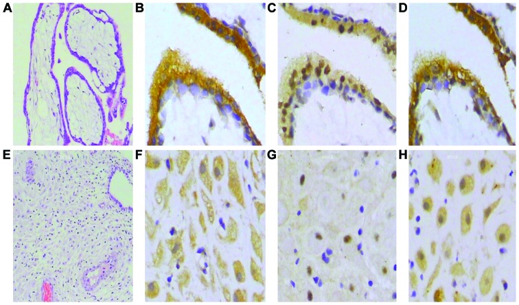
Figure 7.
Photomicrograph of (A) chorionic villi and (B) decidua after hematoxylin and eosin (H&E) staining (×100 magnification). Staining for IDO, SOCS3 and TGF-β revealed their presence in the chorionic villi (B-D) and decidua (E-G), respectively (×400 magnification). Images were taken from the same chorionic villi (A-D) and (E-H) decidua sample, respectively. Staining of human first trimester chorionic villi was localized to cytotrophoblasts (B-D). IDO (B-F) and TGF-β (D-H) were present in the cytoplasm, but SOCS3 was present in both the cytoplasm and nucleus (C-G).

