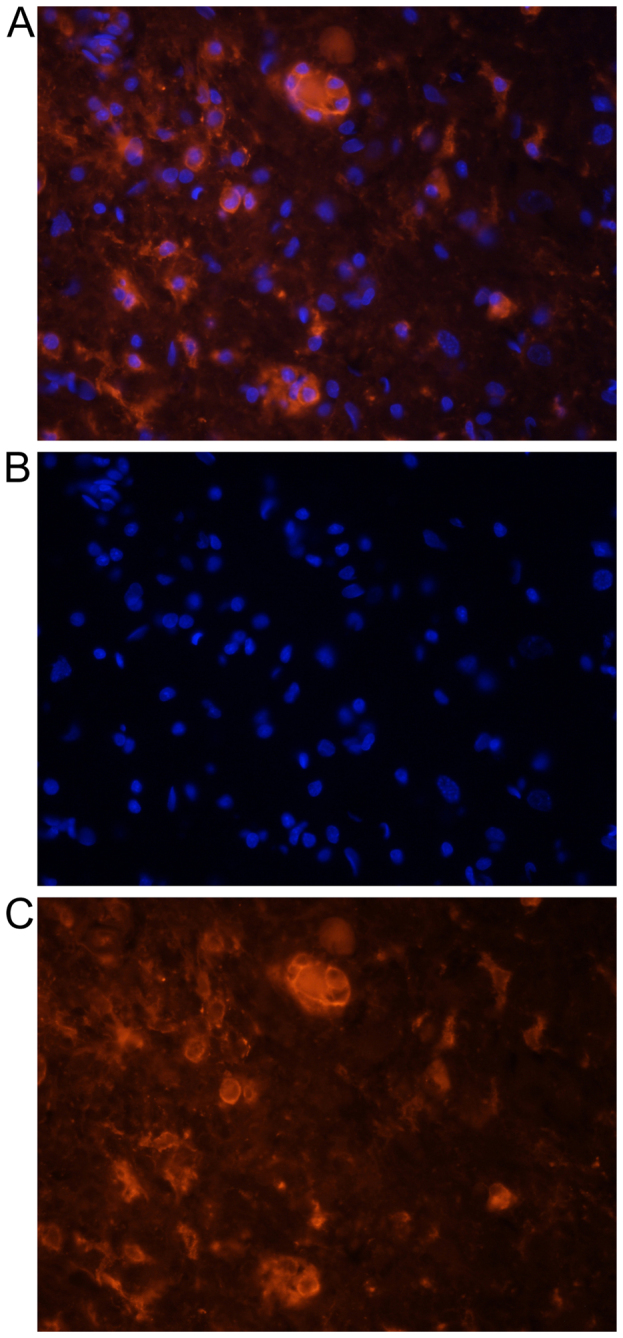
Figure 2.

Representative photographs demonstrating DiI-labeled microglial cells one day after transplantation. (A) Double labeling (DiI and DAPI), (B) DAPI-labeled nuclei and (C) DiI-labeled cytoplasm of microglia. Cells demonstrated a typically round morphology in the nearest vicinity of the main lesion. Microsphere or tumor like formation of these cells is observed. Magnification, ×200. DiI, 1,1′-Dioctadecyl-3,3,3′,3′-Tetramethylindocarbocyanine Perchlorate; DAPI, 4,6-diamidino-2-phenylindole.
