Figure 3.
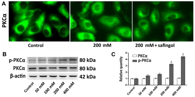
Alterations in the expression of PKCα following ethanol treatment. PKCα expression was detected by immunofluorescence staining and western blotting analyses. (A) Representative immunofluorescence staining images of PKCα following 48 h treatment with 200 mM ethanol or 200 mM ethanol plus safingol (magnification, ×400). (B) Western blotting of PKCα and p-PKCα expression following treatment with different concentrations of ethanol for 48 h. (C) Quantitative analysis of PKCα and p-PKCα protein expression. The results (n=5) are presented as the mean ± standard deviation. *P<0.05 vs. control. p-PKCα, phosphorylated-protein kinase Cα.

