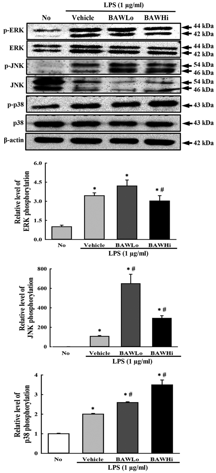
Figure 4.

Expression of three members of the MAP kinase signaling pathway. Nitrocellulose membranes containing 30 µg of total protein from RAW264.7 cells were incubated with antibodies specific to p-ERK, ERK, p-JNK, JNK, p38, p-p38, and β-actin, followed by horseradish peroxidase-conjugated goat anti-rabbit IgG. After the intensity of each band was determined by using an imaging densitometer, the relative levels of the six proteins were calculated based on the intensity of actin protein. The data represent the means ± SD of three replicates. *P<0.05 compared to the No-treated group. #P<0.05 compared to the Vehicle+LPS-treated group.
