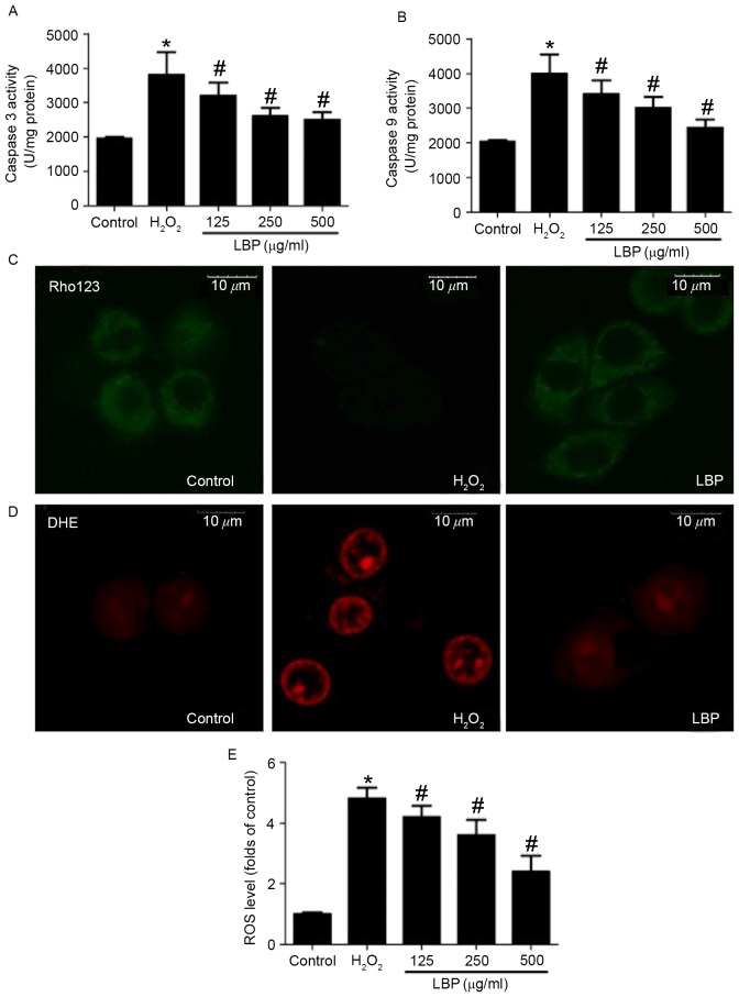
Figure 2.
Effect of LBP on the H2O2-induced mitochondrial apoptotic pathway. PC12 cells were incubated with 400 µM H2O2 in the presence or absence of 125, 250 or 500 µg/ml LBP for 24 h. (A) Caspase-3 and (B) −9 activities were measured using commercial kits and expressed as U/mg protein. (C) PC12 cells were incubated with 400 µM H2O2 in the presence or absence of 500 µg/ml LBP for 24 h. Mitochondrial membrane potential was detected using Rho123 staining. Representative images are presented. ROS levels were determined by DHE and DCFH-DA staining. (D) For DHE staining, a representative image is presented. (E) For DCFH-DA staining, fluorescence was analyzed by flow cytometry and the results are presented as folds of control. *P<0.05 vs. control; #P<0.05 vs. H2O2 treatment. LBP, Lycium barbarum polysaccharide; ROS, reactive oxygen species; DCFH-DA, 7-Dichlorodihydrofluorescein-diacetate; DHE, dihydroethidium; Rho123, Rhodamine 123.

