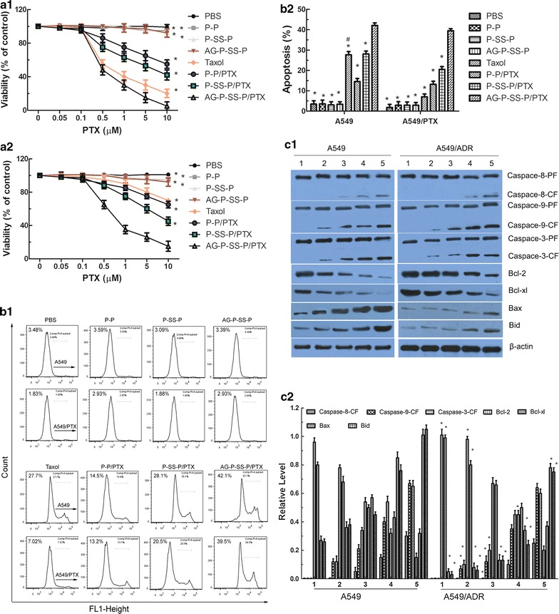
Fig. 6.

a Viability of A549 (a1) and A549/ADR (a2) cells cultured with PTX-loaded nanomicelles in comparison with that of Taxol at the same PTX dose for 48 h. b Cell apoptosis rate detected by flow cytometry. A549 and A549/ADR cells were treated with different formulations that contained a total PTX concentration of 10 µM for 24 h. c Proteins involved in the apoptosis signaling pathways in A549 and A549/ADR cells as determined by Western blotting (c1). (1) Control (PBS); (2) Taxol; (3) P-P/PTX; (4) P-SS-P/PTX; and (5) AG-P-SS-P/PTX nanomicelles. Activity ratios of caspase-3 and caspase-9 and expression ratios of the pro-apoptotic proteins Bax and Bid and the anti-apoptotic proteins Bcl-2 and Bcl-xl in A549 and A549/ADR cells after incubation with the various formulations. β-actin was also assessed by Western blotting. All protein levels were quantified densitometrically and normalized to β-actin (c2). All data are presented as the means ± standard deviations (n = 3); (1) image of western blot; (2) grey level of western blot. *P < 0.05, compared with AG-P-SS-P/PTX nanomicelles. #P < 0.05, compared with A549 cells
